lumbar spine mri anatomy
Myelography (Myelogram) Video: Diagnostic Procedure we have 9 Images about Myelography (Myelogram) Video: Diagnostic Procedure like MRI lumbar spine sagittal cross sectional anatomy image 5 | Anatomy, The Radiology Assistant : Spine - Lumbar Disc Herniation and also Empty thecal sac sign | Image | Radiopaedia.org. Read more:
Myelography (Myelogram) Video: Diagnostic Procedure
 www.spine-health.com
cpt myelogram myelography procedure ligament trapeziectomy reconstruction
The Radiology Assistant : Spine - Lumbar Disc Herniation
 radiologyassistant.nl
lumbar disc herniation nerve compression approach radiology radiologyassistant
MRI Lumbar Spine Sagittal Cross Sectional Anatomy Image 5 | Anatomy
 www.pinterest.com
spine mri lumbar anatomy sagittal cross sectional scans vertebrae disc section intervertebral pelvic use thoracic center explain
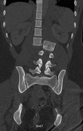
Lumbar Spine Dislocation - Radiology At St. Vincent's University Hospital
 www.svuhradiology.ie
spine lumbar dislocation ct l3 bone radiology lateral left vertebra coronal st
Lumbosacral Plexus (Nerves) | ClipArt ETC
nerves plexus lumbosacral etc clipart usf edu
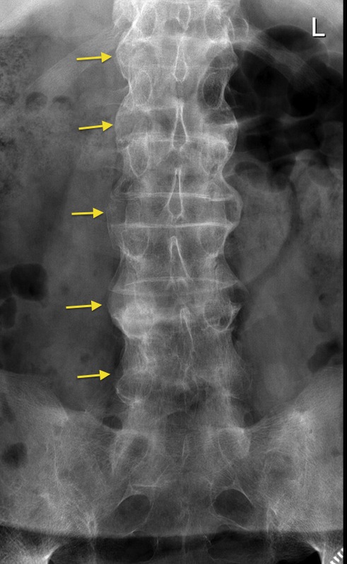
Bamboo Spine Of Ankylosing Spondylitis - Radiology At St. Vincent's
 www.svuhradiology.ie
spondylitis ankylosing spine bamboo radiology case study svuhradiology ie
Empty Thecal Sac Sign | Image | Radiopaedia.org
 radiopaedia.org
thecal axial arachnoid arachnoiditis radiopaedia adhesive nerve
NORMAL AREAS OF SPINAL ENHANCEMENT ON MRI - Radedasia
 radedasia.com
mri intradural spine epidural vein enlargement radiology hypotension anestesia radedasia craniospinal neuroradiology ajnr
Low Back Pain - Medical Clinics
 www.medical.theclinics.com
scoliosis lumbar spine medical mild fig spondylosis pain low hi res theclinics
Lumbosacral plexus (nerves). Normal areas of spinal enhancement on mri. Empty thecal sac sign