malleus incus stapes
Myringostapedopexy - Tympanic Membrane - Mussen Healthcare we have 9 Pictures about Myringostapedopexy - Tympanic Membrane - Mussen Healthcare like Real Human Ear Ossicles - Malleus Incus Stapes - Bones – 6 Brains, Ear Bone Fossils Expected To Yield Key Clues About Human Evolution and also Human Anatomy Lab: Ear Models. Read more:
Myringostapedopexy - Tympanic Membrane - Mussen Healthcare
 www.mussenhealth.us
tympanic membrane cholesteatoma ear surgery retraction pocket through inside tympanoplasty mussen healthcare left figure derick
Process Of Hearing
.PNG) www.sliderbase.com
www.sliderbase.com
hearing process
The Radiology Assistant : Temporal Bone - Anatomy 2.0
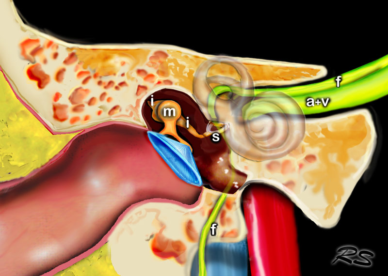 radiologyassistant.nl
bone temporal anatomy ear tympanic radiology middle assistant cavity antrum radiologyassistant nl
The Radiology Assistant : Temporal Bone - Anatomy 2.0
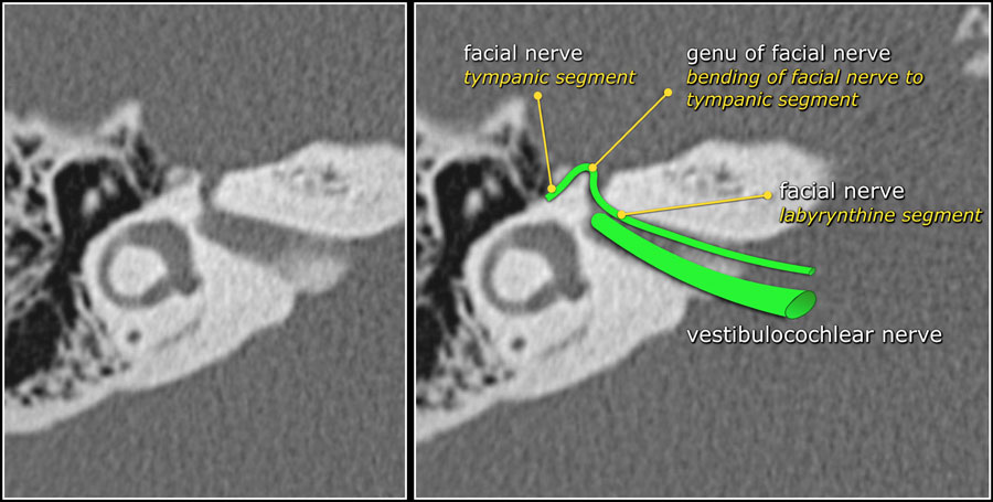 radiologyassistant.nl
temporal bone anatomy nerve facial canal segment radiology labyrinthine auditory internal right petrous geniculate axis ganglion angles sharply nearly coming
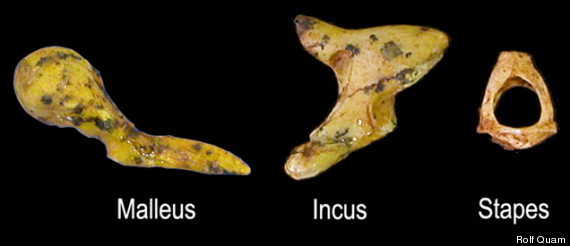
Ear Bone Fossils Expected To Yield Key Clues About Human Evolution
 www.huffingtonpost.com
ear bones human bone three hammer stirrup anvil fossils evolution paranthropus robustus ears malleus incus yield clues expected key stapes
Superior View Of Right Petrous Portion Of Temporal Bone | Neuroanatomy
 www.neurosurgicalatlas.com
petrous correlation neurosurgicalatlas
Real Human Ear Ossicles - Malleus Incus Stapes - Bones – 6 Brains
 6-brains.myshopify.com
ear malleus real ossicles incus stapes bones human
Human Anatomy Lab: Ear Models
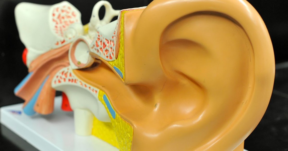 humananatomylab.blogspot.com
ear anatomy human lab models utricle duct canals vestibule
Middle Fossa Exposure Of The Temporal Bone | Neuroanatomy | The
 www.neurosurgicalatlas.com
temporal bone fossa exposure middle neurosurgicalatlas correlation surgical
Middle fossa exposure of the temporal bone. The radiology assistant : temporal bone. Temporal bone anatomy nerve facial canal segment radiology labyrinthine auditory internal right petrous geniculate axis ganglion angles sharply nearly coming